|
Page
6 |
A&P
1
Lab Manual |
If you disable the "Active Content" in your browser you may not
be able to view the animations or videos supplied in this lab.
If prompted you should "Allow Blocked Content". |
|
|
|
|
|
Lab 3
Tissues and Skin |
 |
|
|
Access each of the listed
documents above and print them off. When you
submit your lab report you will need to
compile all of the documents listed above,
stapled together in the order listed in the
table above. Sketches must be performed free
hand (not traced or copy and pasted).
Sketches must be performed using the printed
links as given above. You are not allowed to
perform the sketches on blank sheets of
paper or lined sheets of paper. Sketches
performed without using these forms above
will not be accepted. You can use the
MS WORD links to access the questions,
tables and charts in order to input your
values or answers electronically and then
print them off when finished to include with
your lab report. Alternatively you can print
the questions, tables and charts forms out
and input your values or answers by hand.
The PDF file format will not allow you to
input values or answers electronically.
Please collate and order the pages in your
lab report in the order they are listed in
the table above. The cover page is only
available using the PDF file format. |
1)
Tissues
A tissue
is a collection of cells specialized for a particular common
function. The amount of intercellular material varies
significantly in different tissue types, from being very
dispersed to forming the bulk of the tissue. Microscopic
anatomists have recognized that all of the cells of the
human body can be classified into four basic tissues.
Criteria for this organization are based on similarity in
cell function, appearance, origin and products formed.
Epithelial tissue
This is the covering or protective layer on most free
surfaces or cavities.
Connective tissue
This is the tissue that serves as an anchor for the
other tissues. In addition this tissue has some forms
that are specialized.
Muscular tissue
This tissue has the ability to generate large amounts of
force and cause its length to decrease. This is the
tissue that allows mobility.
Nervous tissue
This tissue is the information gathering, decision
making, and action causing network of the body.
A) EPITHELIAL
TISSUE
Epithelial tissues form sheet-like coverings or linings
which can be very thick or in other cases very thin. The
adherent nature of the epithelial cells allows the
formation of continuous sheets of tissue. Most free
surfaces of the body are covered by epithelial tissues.
Epithelial tissues are devoid of blood vessels, but
often rich in nerve endings.
Epithelial types are classified for the most part by the
shape of the cells. The types of epithelia found in a given
location is a reflection of its function. Areas of wear and
tear require a thick epithelium, like those of the epidermis
and oral cavity, while the lining of small blood vessels do
not. Areas of thin epithelium may facilitate the transfer of
materials in absorption and secretion. Areas, like
epidermis, exposed to drying factors are keratinized. Areas,
in the oral region, need not to be keratinized due to the
continual moisture that is present.
SKETCH 1
**Using
the images provided below, view and sketch the
following epithelial tissues. You can use the forms given
below to produce your sketches.
Tissue
Sketch Form (MS WORD)
Tissue Sketch
Form (PDF)
|
Simple epithelium
Simple
squamous
Simple
cuboidal
Simple
columnar
Pseudostratified epithelium
Ciliated epithelium
Stratified epithelium
Stratified squamous epithelium

Figure 3.1 Simple Squamous (alveoli from lungs)
|

Figure 3.2 Epithelium Tissue Types |

Figure 3.3 Simple Cuboidal
(kidney tubule)
|

Figure 3.4
Simple Columnar Epithelium (small intestine)
|
|

Figure 3.5
Pseudostratified Epithelium (trachea)
|

Figure 3.6
Stratified Squamous Epithelium (esophagus lining)
|
B)
CONNECTIVE
TISSUE
Connective tissue, in contrast to epithelial tissue,
contains relatively few cells and a large amount of
intercellular material. Connective tissue is the most
abundant and widely distributed of the tissue types. The
extracellular material consists of many types of non-living
substances which is produced and exuded by the connective
tissue cells. Collectively the material is called the
extracellular matrix. The matrix provides the characteristic
strength of connective tissues. Connective tissue may be
classified as:
Dense fibrous
-
found in tendons and ligaments
Loose areolar
-
found usually as packing around organs
Adipose
-
commonly called fat
Cartilage
- usually found capping long bone
(hyaline)
Bone
-
makes up the skeletal system
Blood
-
type of tissue bathed in plasma and includes numerous
different types of cells
SKETCH 2
**Using
the images provided below, view and sketch the
following connective tissues:
Dense
fibrous,
Loose areolar, Adipose, Cartilage, Bone, Blood.
(You can use the forms given below to produce your sketches)
Connective Tissue
Sketch Form (MS WORD)
Connective Tissue Sketch
Form (PDF)

Figure 3.7
Dense Fibrous Connective Tissue (tendon)
|

Figure 3.8 Loose Areolar
Tissue
|

Figure 3.9 Adipose Tissue
|

Figure 3.10 Bone Tissue
|
|

Figure 3.11
Blood Cells: Red blood cells (left), White blood cells
(right)
|
|

Figure 3.12 Cartilage Tissue
|
C)
MUSCLE TISSUE
Muscle
tissue is highly specialized to contract in order to produce
movement of some body parts. Muscle cells tend to be quite
elongated, providing a long axis for contraction. For the
most part muscle tissue has the singular capability of being
able to contract. The three
basic types of muscle tissue are as follows:
Skeletal
- voluntary, non-branching,
striated, multinucleated, attached to bone
Cardiac
- involuntary, branching , striated, single nucleus,
heart muscle
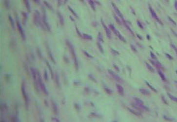
Smooth
- involuntary, spindled, non-striated, single nucleus,
walls of hollow organs
SKETCH 3
**Using
the images provided below, sketch the following
muscle tissues:
Skeletal muscle, Cardiac muscle,
Smooth muscle
(You can use the forms given
below to produce your sketches)
Muscle Tissue
Sketch Form (MS WORD)
Muscle Tissue Sketch
Form (PDF)
|

Figure 3.13 Skeletal Muscle Tissue
|

Figure 3.14 Cardiac Muscle Tissue
|
|

Figure 3.15 Smooth Muscle Tissue
|
D)
NERVOUS TISSUE
The
structure of nerve tissue is markedly different from that of
all other body cells. All of the cells have a nucleus. These
cells are designed for being very efficient at carrying a
nerve impulse.
Neurons
- conduct waves of excitation
SKETCH 4
**Using
the images provided below, sketch the following
nervous tissue: Neruons.
You can use
the forms given below to produce your sketches.
Nerve Tissue
Sketch Form (MS WORD)
Nerve Tissue Sketch
Form (PDF)
|

Figure 3.16 Neurons in the
Cerebrum of the Brain
|

Figure 3.17 Neuron
|
Click here to view a tutorial on Tissue Types
Tissue
Types Tutorial
Click here to view a tutorial on Muscle and Connective
Tissue
Muscle and Connective Tissue Tutorial
Click here to view a tutorial on Nervous and Epithelial
Tissues
Nervous and Epithelial Tissue Tutorial
QUESTIONS
**Questions on Tissue
Function
1)
List two functions of the skin
2)
List two
characteristics of epithelial tissue
3)
Name two
organs where we would find striated muscle
4)
Concentric rings are found in what type of tissue?
5)
Most of
the volume of adipose tissue cells is what molecule?
6)
List an
organ where we would find the following tissue types:
QUESTIONS
Histology Questions
Answers the questions indicated at the HISTOLOGY QUIZ
link given below. List your answers 1-14

Histology Questions Forms (MS WORD)
Histology Questions Forms
(PDF)
2) Skin
Anatomy
The
integument is often considered an organ system because of its
extent and complexity. The skin has many functions, most
concerned with protection. The skin has two distinct regions,
the superficial epidermis and the underlying dermis.
Immediately below the dermis is the hypodermis. There are a
number of accessory organs and tissue types in the skin as well.
SKETCH 5
**Using
the images of the slides of human skin and the skin models
given below, construct a series of sketches which would
enable you to label and identify the following:
Stratum corneum, Stratum granulosum, Stratum
basale, Epidermis, Stratum spinosum, Dermis, Hypodermis,
Sweat gland,
Papillary layer, Reticular layer, Hair
follicle, Sebaceous gland, Arrector pilli muscle,
Hair shaft
Click here to explore
an interactive study on the integumentary system
INTEGUMENTARY SYSTEM
Click
here to explore an interactive study on the skin layers
SKIN LAYERS
Click
here to explore an interactive study on skin cells
SKIN CELLS
Click
here to explore an interactive study on accessory organs of the
skin
ACCESSORY ORGANS OF THE SKIN
Click
here to explore an interactive study of sweat glands and hair
follicles
SWEAT GLANDS AND HAIR FOLLICLES
Click here to see a
movie on skin healing and regeneration of skin
SKIN HEALING
Dissection of Cadaver Skin
Use the link below to access the video. Once
there click on the image to view the movie. The
dissection begins at about the 2 minute mark.
Human Cadaver Skin Dissection

Figure 3.18 Skin Model
|

Figure 3.19 Skin Model
|

Figure 3.20 Skin Model
|

Figure 3.21 Microscopic Cross
Section of Skin
|

Figure 3.22 Microscopic Cross
Section of Skin
|

Figure 3.23 Microscopic Cross
Section of Skin
|

Figure 3.24 Microscopic Cross
Section of Skin
|
SKIN CANCER
Skin cancer is the most
common cancer in the U.S. Approximately 2 million people are
diagnosed annually with skin cancer. There are more cases of
skin cancer diagnosed than lung, colon, breast and prostate
cancer combined. This means that approximately 1 in 5 people
will be diagnosed with skin cancer in their lifetime.
The number one risk factor for skin
cancer is UV radiation exposure.
The most common source of UV
radiation is sunlight.
In fact, most people have
experienced more than 50% of the recommended lifetime UV dose by
the time they are 20 years old.
UV radiation exposure can also occur
in tanning devices.
In fact, a single use of a
tanning device increases the chance of developing skin cancer by
20%.
According to the American
Cancer Society, those who begin indoor tanning before the age of
35 have an 87% increase in their risk of developing melanoma.
Other risk factors beside UV radiation
exposure that increase risk of skin cancer include:
Fair Skin
Living in a sunny
environment
Moles
Skin lesions
Family History
Compromised immune system
Radiation exposure
Chemical
exposure
Types of Skin Cancer
Skin cancer is most simply abnormal
growth of epithelial cells, and is most commonly found in areas
of skin with high sun exposure.
UV radiation causes mutations
in the DNA of skin cells, and when these mutations occur in
parts of the DNA that control cell growth, uncontrolled cell
growth occurs.
There are three main types of
skin cancer separated by the type of epithelial cells that they
develop.
Epithelial cells divide
quickly, and many skin cancers can develop quickly depending on
their location.
Luckily, nearly all forms of
skin cancer are easily treatable through surgical excision if
detected, diagnosed, and treated early.
The following table describes
the three most common types of skin cancer.
|
Skin Cancer
Type
|
Cells Where Develops
|
Description
|
|
Squamous Cell Carcinoma

|
Squamous cells
primarily make up the cells found in the
epidermis.
|
20% of all skin cancers are squamous cell
carcinoma.
This cancer is more aggressive than basal
cell carcinoma, but easily treated when found
early.
Squamous cell carcinoma will look like a
red, scaly bump or nodule and is most commonly
found on the face.
It can easily spread to other parts of
the body, and is more common in individuals with
fair skin.
|
|
Basal Cell
Carcinoma

|
Basal cells
lie just below the epidermis and create the
basement layer that nourishes the epidermis.
|
The most common type
of skin cancer, making up 75% of all skin
cancers, is basal cell carcinoma.
This cancer grows very slowly and looks
like shiny, waxy bumps or nodules on the skin.
It is most commonly found on areas of the
body with high sun exposure like the head, arms,
legs, and face.
|
|
Melanoma

|
Melanocytes
are spread throughout the skin and are
responsible for producing the pigment that
creates skin color.
|
Melanoma is the least common type of cancer, but
accounts for more than 75% of all deaths caused
by skin cancer.
This cancer most commonly starts as a
mole that becomes cancerous and appears as a
large brown spot with irregular borders.
Due to the pigment produced by
melanocytes, it will often appear as if the mole
is growing.
Melanoma is most commonly found on the
head, neck, or trunk.
|
|
Patient
Analysis
Finding suspicious moles or
skin cancer early is the key to treating skin cancer
successfully.
Examining
yourself is usually the first step in detecting skin
cancer. You are a family physician and you have a few
patients coming in to have their moles observed.
For each patient use
the ABCD chart to identify whether the mole is
suspicious.
Write a summary for
each of the patients provided, answering the following:
1) Is there a possibility the mole
is cancerous and why or why not?
2)
What is your advice to the patient for this mole? (none,
proper sun care, removal, etc.)
|
 |
END LAB 3
|